jeff orchard’s brain

I’ve had my “head examined” on more than one occasion. In particular, I have had a couple magnetic resonance imaging (MRI) scans done of my head. One at UBC hospital, from which I acquired 18 sagital slices and 18 axial slices, and one at BC Children’s Hospital (129 sagital slices).
These MRI scans are T1 weighted, which means that bone and air are black, while bone marrow and other soft tissues are gray or white. Blood that is just arriving to the head will also appear black.
The best way to get your hands on my MRI data is to download ImageJ. My dataset is one of the samples that you can download in the “File” menu.
Download a TIFF file of my T1 head dataset (4MB)
Download a gnuzipped file of my raw T1 head dataset
(256x256x129, unsigned integer, 16-bit, little endian, 5.5MB)


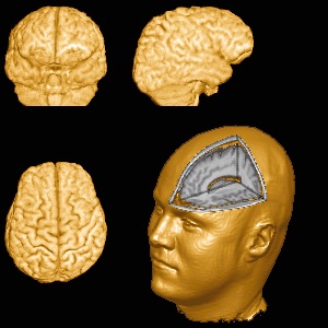

3d renderings
These were produced by Chris Rorden , the creator of MRIcro
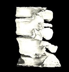
This is a CT scan of my lumbar spine. Some of the structures are wrong because not all slices were acquired in the same plane.
See my brain through my head.
Animation of my rotating brain (2.9MB GIF)
